
Click image to see more details
Product Info Summary
| SKU: | PB9234 |
|---|---|
| Size: | 100 μg/vial |
| Reactive Species: | Human, Mouse, Rat |
| Host: | Rabbit |
| Application: | Flow Cytometry, IF, IHC, ICC, WB |
Customers Who Bought This Also Bought

Product info
Product Name
Anti-ZO1 tight junction protein/TJP1 Antibody Picoband™
View all Tight Junction Protein 1 Antibodies
SKU/Catalog Number
PB9234
Size
100 μg/vial
Form
Lyophilized
Description
Boster Bio Anti-ZO1 tight junction protein/TJP1 Antibody Picoband™ catalog # PB9234. Tested in Flow Cytometry, IF, IHC, ICC, WB applications. This antibody reacts with Human, Mouse, Rat.
Storage & Handling
Store at -20˚C for one year from date of receipt. After reconstitution, at 4˚C for one month. It can also be aliquotted and stored frozen at -20˚C for six months. Avoid repeated freeze-thaw cycles.
Cite This Product
Anti-ZO1 tight junction protein/TJP1 Antibody Picoband™ (Boster Biological Technology, Pleasanton CA, USA, Catalog # PB9234)
Host
Rabbit
Contents
Each vial contains 5mg BSA, 0.9mg NaCl, 0.2mg Na2HPO4, 0.05mg NaN3.
Clonality
Polyclonal
Isotype
Rabbit IgG
Immunogen
E.coli-derived human TJP1 recombinant protein (Position: H1178-F1527). Human TJP1 shares 82% amino acid (aa) sequence identity with mouse TJP1.
*Blocking peptide can be purchased. Costs vary based on immunogen length. Contact us for pricing.
Cross-reactivity
No cross-reactivity with other proteins
Reactive Species
PB9234 is reactive to TJP1 in Human, Mouse, Rat
Applications
PB9234 is guaranteed for Flow Cytometry, IF, IHC, ICC, WB Boster Guarantee
Observed Molecular Weight
220 kDa
Calculated molecular weight
195.459kDa
Background of Tight Junction Protein 1
Tight junction protein ZO-1 is a protein that in humans is encoded by the TJP1 gene. It is mapped to 15q13.1. This gene encodes a protein located on a cytoplasmic membrane surface of intercellular tight junctions. The encoded protein may be involved in signal transduction at cell–cell junctions. It has been found that injected CagA associates with the epithelial tight-junction scaffolding protein TJP1 and the transmembrane protein junctional adhesion molecule, causing an ectopic assembly of tight junction components at sites of bacterial attachment, and altering the composition and function of the apical-junctional complex.
Antibody Validation
Boster validates all antibodies on WB, IHC, ICC, Immunofluorescence, and ELISA with known positive control and negative samples to ensure specificity and high affinity, including thorough antibody incubations.
Innovating Scientists Reward
If you are the first to review this product, or if you have results for a special sample, species or application this product is not validated in, share your results with us and receive product credits you can use towards any Boster products! Applicable to all scientists worldwide.
Submit A Review
Assay dilution & Images
Reconsitution
Add 0.2ml of distilled water will yield a concentration of 500ug/ml.
Assay Dilutions Recommendation
The recommendations below provide a starting point for assay optimization. The actual working concentration varies and should be decided by the user.
Western blot, 0.1-0.5μg/ml, Human, Mouse, Rat
Immunohistochemistry (Paraffin-embedded Section), 0.5-1μg/ml, Human, By Heat
Immunocytochemistry/Immunofluorescence, 2μg/ml, Human
Flow Cytometry, 1-3μg/1x106 cells, Human
Validation Images & Assay Conditions
Click image to see more details
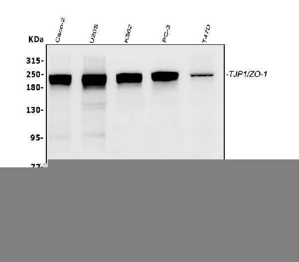
Figure 1. Western blot analysis of TJP1 using anti-TJP1 antibody (PB9234).
Electrophoresis was performed on a 5-20% SDS-PAGE gel at 70V (Stacking gel) / 90V (Resolving gel) for 2-3 hours. The sample well of each lane was loaded with 30 ug of sample under reducing conditions.
Lane 1: human PC-3 whole cell lysates,
Lane 2: human CACO-2 whole cell lysates,
Lane 3: human COLO320 whole cell lysates,
Lane 4: rat testis tissue lysates,
Lane 5: mouse testis tissue lysates.
After electrophoresis, proteins were transferred to a nitrocellulose membrane at 150 mA for 50-90 minutes. Blocked the membrane with 5% non-fat milk/TBS for 1.5 hour at RT. The membrane was incubated with rabbit anti-TJP1 antigen affinity purified polyclonal antibody (Catalog # PB9234) at 0.5 μg/mL overnight at 4°C, then washed with TBS-0.1%Tween 3 times with 5 minutes each and probed with a goat anti-rabbit IgG-HRP secondary antibody at a dilution of 1:5000 for 1.5 hour at RT. The signal is developed using an Enhanced Chemiluminescent detection (ECL) kit (Catalog # EK1002) with Tanon 5200 system. A specific band was detected for TJP1 at approximately 220 kDa. The expected band size for TJP1 is at 185 kDa.

Click image to see more details
Figure 2. IHC analysis of TJP1 using anti-TJP1 antibody (PB9234).
TJP1 was detected in paraffin-embedded section of human intestinal cancer tissue. Heat mediated antigen retrieval was performed in citrate buffer (pH6, epitope retrieval solution) for 20 mins. The tissue section was blocked with 10% goat serum. The tissue section was then incubated with 1μg/ml rabbit anti-TJP1 Antibody (PB9234) overnight at 4°C. Biotinylated goat anti-rabbit IgG was used as secondary antibody and incubated for 30 minutes at 37°C. The tissue section was developed using Strepavidin-Biotin-Complex (SABC)(Catalog # SA1022) with DAB as the chromogen.

Click image to see more details
Figure 3. IF analysis of TJP1 using anti-TJP1 antibody (PB9234).
TJP1 was detected in immunocytochemical section of A431 cells. Enzyme antigen retrieval was performed using IHC enzyme antigen retrieval reagent (AR0022) for 15 mins. The cells were blocked with 10% goat serum. And then incubated with 2μg/mL rabbit anti-TJP1 Antibody (PB9234) overnight at 4°C. DyLight®488 Conjugated Goat Anti-Rabbit IgG (BA1127) was used as secondary antibody at 1:100 dilution and incubated for 30 minutes at 37°C. The section was counterstained with DAPI. Visualize using a fluorescence microscope and filter sets appropriate for the label used.

Click image to see more details
Figure 4. Flow Cytometry analysis of K562 cells using anti-TJP1 antibody (PB9234).
Overlay histogram showing K562 cells stained with PB9234 (Blue line).The cells were blocked with 10% normal goat serum. And then incubated with rabbit anti-TJP1 Antibody (PB9234,1μg/1x106 cells) for 30 min at 20°C. DyLight®488 conjugated goat anti-rabbit IgG (BA1127, 5-10μg/1x106 cells) was used as secondary antibody for 30 minutes at 20°C. Isotype control antibody (Green line) was rabbit IgG (1μg/1x106) used under the same conditions. Unlabelled sample (Red line) was also used as a control.
Protein Target Info & Infographic
Gene/Protein Information For TJP1 (Source: Uniprot.org, NCBI)
Gene Name
TJP1
Full Name
Tight junction protein ZO-1
Weight
195.459kDa
Superfamily
MAGUK family
Alternative Names
DKFZp686M05161; MGC133289; tight junction protein 1 (zona occludens 1); Tight junction protein 1; tight junction protein ZO-1; TJP1; ZO1; ZO-1; zona occludens 1; Zona occludens protein 1; zonula occludens 1 protein; Zonula occludens protein 1 TJP1 ZO-1 tight junction protein 1 tight junction protein ZO-1|zona occludens 1|zonula occludens 1 protein
*If product is indicated to react with multiple species, protein info is based on the gene entry specified above in "Species".For more info on TJP1, check out the TJP1 Infographic

We have 30,000+ of these available, one for each gene! Check them out.
In this infographic, you will see the following information for TJP1: database IDs, superfamily, protein function, synonyms, molecular weight, chromosomal locations, tissues of expression, subcellular locations, post-translational modifications, and related diseases, research areas & pathways. If you want to see more information included, or would like to contribute to it and be acknowledged, please contact [email protected].
Specific Publications For Anti-ZO1 tight junction protein/TJP1 Antibody Picoband™ (PB9234)
Hello CJ!
PB9234 has been cited in 32 publications:
*The publications in this section are manually curated by our staff scientists. They may differ from Bioz's machine gathered results. Both are accurate. If you find a publication citing this product but is missing from this list, please let us know we will issue you a thank-you coupon.
Protective effect of salvianolic acid B on NASH rat liver through restoring intestinal mucosal barrier function
Dual Receptor Recognizing Cell Penetrating Peptide for Selective Targeting, Efficient Intratumoral Diffusion and Synthesized Anti-Glioma Therapy
Piperazine ferulate prevents high‑glucose‑induced filtration barrier injury of glomerular endothelial cells
The Regulatory Effects of Licochalcone A on the Intestinal Epithelium and Gut Microbiota in Murine Colitis
Prevention and Alleviation of Dextran Sulfate Sodium Salt-Induced Inflammatory Bowel Disease in Mice With Bacillus subtilis-Fermented Milk via Inhibition of the Inflammatory Responses and Regulation of the Intestinal Flora
The Neurovascular Protective Effects of Huperzine A on D-Galactose-Induced Inflammatory Damage in the Rat Hippocampus
Vancomycin and ceftriaxone can damage intestinal microbiota and affect the development of the intestinal tract and immune system to different degrees in neonatal mice
Genetically encoded probiotic EcN 1917 alleviates alcohol-induced acute liver injury and restore gut microbiota homeostasis
Bifidobacterium breve ATCC15700 pretreatment prevents alcoholic liver disease through modulating gut microbiota in mice exposed to chronic alcohol intake
Protective effects of acarbose against vascular endothelial dysfunction through inhibiting Nox4/NLRP3 inflammasome pathway in diabetic rats
Recommended Resources
Here are featured tools and databases that you might find useful.
- Boster's Pathways Library
- Protein Databases
- Bioscience Research Protocol Resources
- Data Processing & Analysis Software
- Photo Editing Software
- Scientific Literature Resources
- Research Paper Management Tools
- Molecular Biology Software
- Primer Design Tools
- Bioinformatics Tools
- Phylogenetic Tree Analysis
Customer Reviews
Have you used Anti-ZO1 tight junction protein/TJP1 Antibody Picoband™?
Submit a review and receive an Amazon gift card.
- $30 for a review with an image
Be the first to review Anti-ZO1 tight junction protein/TJP1 Antibody Picoband™
*The first user to submit a review for a product is eligible for Boster's Innovating Scientists Reward, which gives product credits. This is in addition to the gift card reward.
Customer Q&As
Have a question?
Find answers in Q&As, reviews.
Can't find your answer?
Submit your question
5 Customer Q&As for Anti-ZO1 tight junction protein/TJP1 Antibody Picoband™
Question
Is a blocking peptide available for product anti-ZO1 tight junction protein/TJP1 antibody (PB9234)?
Verified Customer
Verified customer
Asked: 2019-11-21
Answer
We do provide the blocking peptide for product anti-ZO1 tight junction protein/TJP1 antibody (PB9234). If you would like to place an order for it please contact [email protected] and make a special request.
Boster Scientific Support
Answered: 2019-11-21
Question
We are currently using anti-ZO1 tight junction protein/TJP1 antibody PB9234 for rat tissue, and we are happy with the WB results. The species of reactivity given in the datasheet says human, mouse, rat. Is it likely that the antibody can work on primate tissues as well?
Verified Customer
Verified customer
Asked: 2019-09-23
Answer
The anti-ZO1 tight junction protein/TJP1 antibody (PB9234) has not been tested for cross reactivity specifically with primate tissues, though there is a good chance of cross reactivity. We have an innovator award program that if you test this antibody and show it works in primate you can get your next antibody for free. Please contact me if I can help you with anything.
Boster Scientific Support
Answered: 2019-09-23
Question
For PB9234's WB image, the customer would like to confirm that the antibody dilution as 0.5ug/ml. Also, the customer wanted to know the detailed transfer conditions for the QC data, is the information on the WB image sufficient or did we have more information?
Verified Customer
Verified customer
Asked: 2019-07-29
Answer
The antibody dilution is 0.5ug/mL. After Electrophoresis, proteins were wet transferred to a Nitrocellulose membrane at 150mA for 90 minutes.
Boster Scientific Support
Answered: 2019-07-29
Question
Our lab want to know about to test anti-ZO1 tight junction protein/TJP1 antibody PB9234 on rat liver for research purposes, then I may be interested in using anti-ZO1 tight junction protein/TJP1 antibody PB9234 for diagnostic purposes as well. Is the antibody suitable for diagnostic purposes?
Verified Customer
Verified customer
Asked: 2018-10-10
Answer
The products we sell, including anti-ZO1 tight junction protein/TJP1 antibody PB9234, are only intended for research use. They would not be suitable for use in diagnostic work. If you have the means to develop a product into diagnostic use, and are interested in collaborating with us and develop our product into an IVD product, please contact us for more discussions.
Boster Scientific Support
Answered: 2018-10-10
Question
Would anti-ZO1 tight junction protein/TJP1 antibody PB9234 work on feline WB with cervix carcinoma erythroleukemia?
Verified Customer
Verified customer
Asked: 2018-01-03
Answer
Our lab technicians have not validated anti-ZO1 tight junction protein/TJP1 antibody PB9234 on feline. You can run a BLAST between feline and the immunogen sequence of anti-ZO1 tight junction protein/TJP1 antibody PB9234 to see if they may cross-react. If the sequence homology is close, then you can perform a pilot test. Keep in mind that since we have not validated feline samples, this use of the antibody is not covered by our guarantee. However we have an innovator award program that if you test this antibody and show it works in feline cervix carcinoma erythroleukemia in WB, you can get your next antibody for free.
Boster Scientific Support
Answered: 2018-01-03
